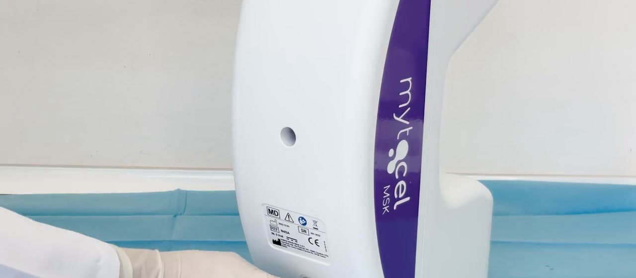
Autologous cartilage micrografting
Mytocel MSK. Prepared from the patient’s own cartilage, at the point of care.
Mytocel MSK® is an autologous cartilage micrografting procedure: the patient’s own auricular cartilage, mechanically disaggregated at the point of care into an injectable micrograft suspension.
- Tissue source
- Auricular cartilage, three 2.5 mm biopsies
- Processing
- Mechanical disaggregation, 80 µm
- Laboratory step
- None
- Session
- One outpatient visit, about 30 minutes
- Devices
- Rigeneracons® RS (Class IIa), N4SA (Class I)
- UK supply
- Actomed, exclusive
How it works
Four stages. One outpatient session.
Sampling, processing and administration in one outpatient session of around 30 minutes.
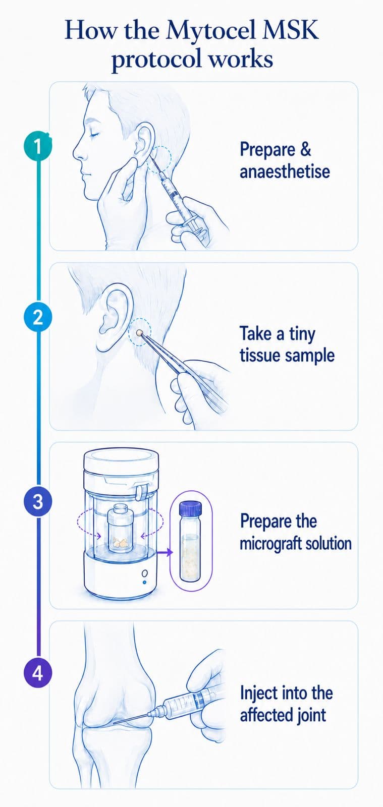
- 01
Prepare
Establish the sterile field and prepare the posterior auricular donor area under local anaesthetic, following the current professional protocol.
- 02
Sample
Take three 2.5 mm punch biopsies from the posterior auricular cartilage using the documented technique. No sutures are required.
- 03
Process
Disaggregate the samples mechanically in Rigeneracons® RS on the N4SA processing unit and collect the AMT® suspension through the calibrated 80 µm filter. No laboratory step.
- 04
Administer
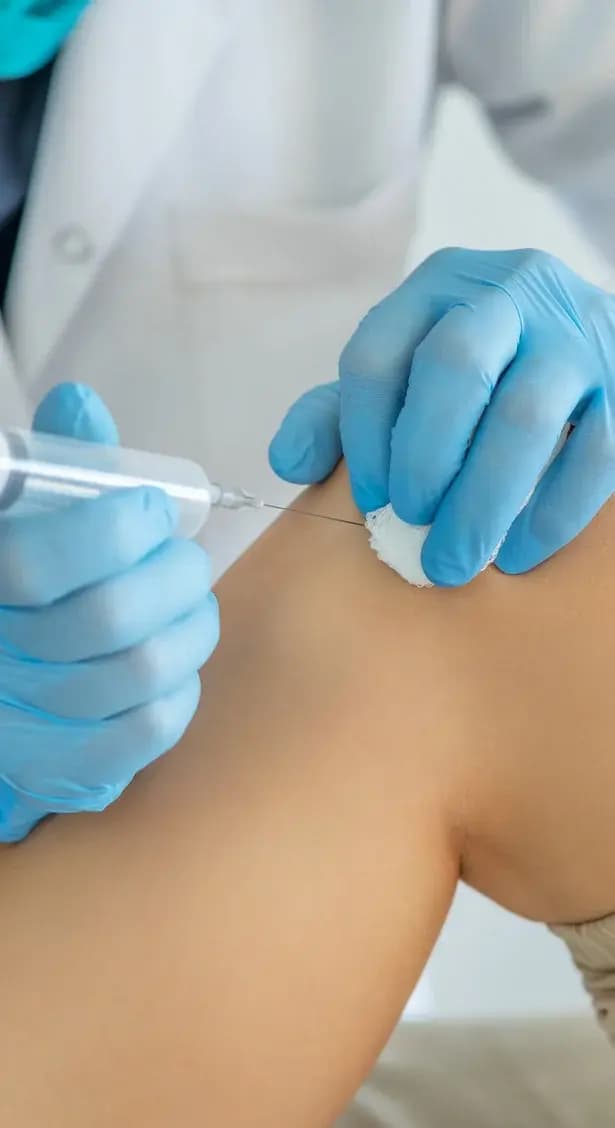
Administer the suspension to the target joint by intra-articular injection within the same session, then complete aftercare and follow-up arrangements.
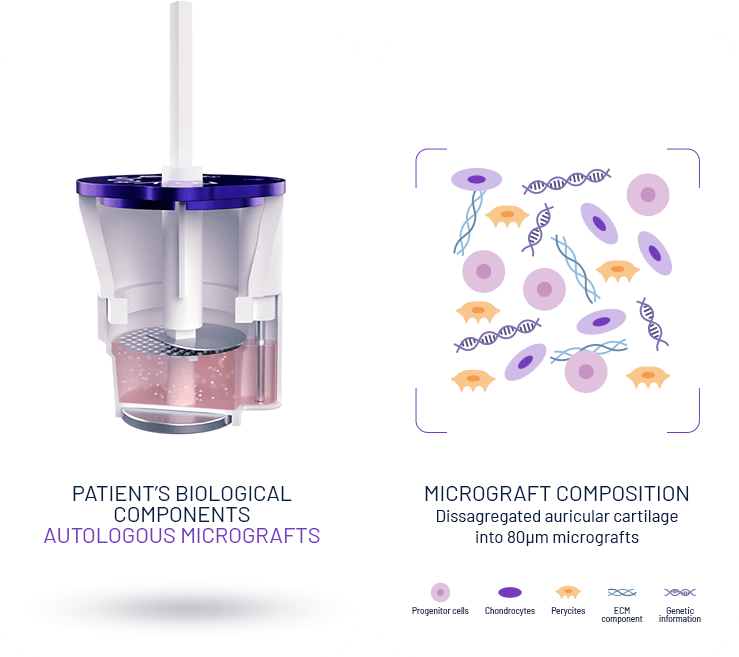
What the suspension contains
A suspension prepared from the patient’s own cartilage.
Characterisation studies describe a heterogeneous suspension of cartilage-derived cells (chondrocytes and progenitor cells), pericytes and extracellular-matrix components from the donor tissue.1
Mechanical disaggregation only: no laboratory expansion, no enzymatic digestion, no culture step.
Micrografts are collected through a calibrated 80 µm filter, the size range associated with viable progenitor-rich fractions in the characterisation work.1
Evidence
Three-year follow-up. Two prospective series. Every study linked in full.
Indications
Documented for early to moderate cartilage disease.
Manufacturer-documented indications: knee osteoarthritis grade I to III (Kellgren-Lawrence), focal cartilage injury or damage, chondromalacia patellae.
Not indicated in grade IV
The system
Two devices, one documented processing cycle.
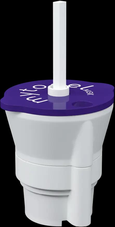
Rigeneracons RS
Disaggregation device
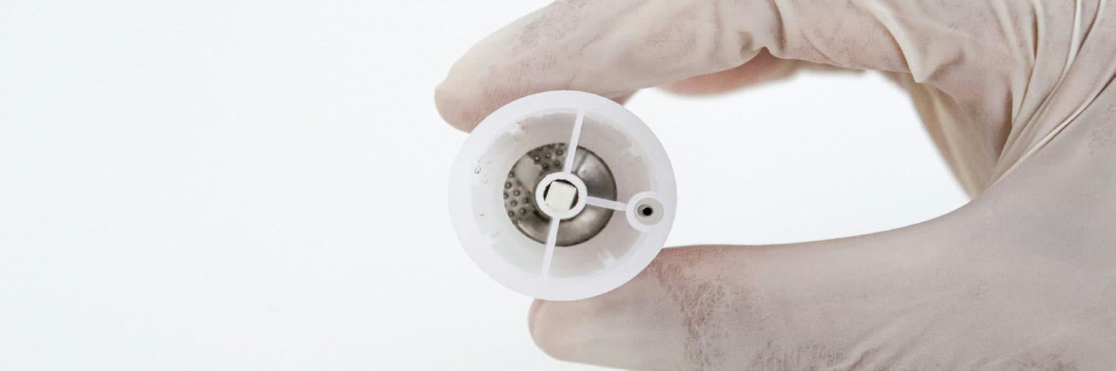
Rigeneracons® RS: sterile, single-use Class IIa medical device, designed to mechanically disaggregate the auricular cartilage sample and collect the micrograft suspension through the calibrated 80 µm filter.
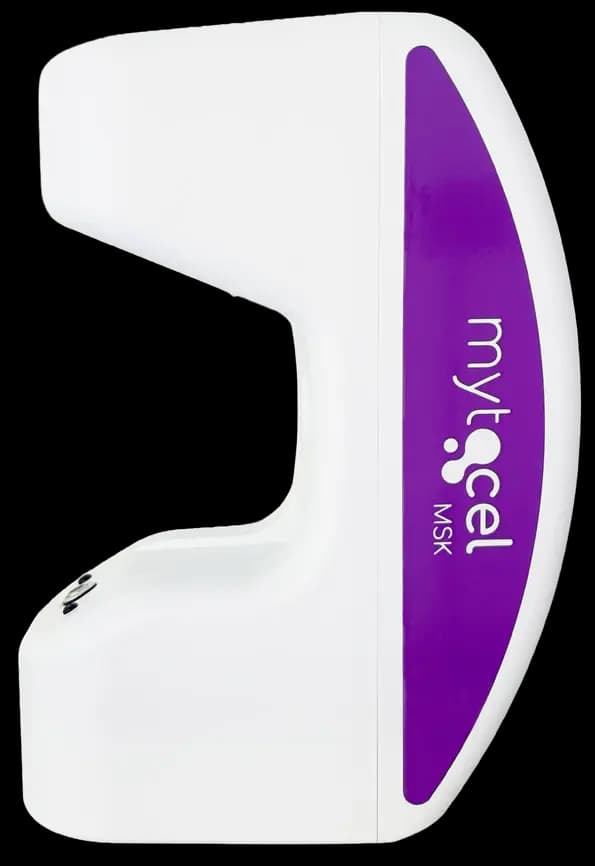
Rigenera N4SA
Processing unit
Rigenera® N4SA: Class I motor-driven processing unit. The reusable unit provides the calibrated rotational impulse for the documented processing cycle.
Rigeneracons® RS: sterile, single-use Class IIa medical device. Rigenera® N4SA: Class I motor-driven processing unit. CE marked; accepted on the Great Britain market under current transitional arrangements. Registered with the MHRA.
Working with Actomed
Exclusive UK supply, structured onboarding.
Actomed is the exclusive UK supplier of Mytocel MSK, holding stock of Rigeneracons® RS and supporting N4SA installation.
Structured onboarding: protocol training, equipment orientation, first-case support and a documented consumables list.
KOOS and feedback templates for structured local outcome collection.
Onboarding in five steps
- 01
Professional discussion
Clinical fit, evidence questions and clinic objectives.
- 02
Pricing and configuration
Reusable equipment, single-use devices and expected activity.
- 03
Training and preparation
Protocol training, equipment orientation, setup and supporting documentation.
- 04
First-case pathway
Agree practical first-case support and readiness before clinical use.
- 05
Ongoing supply
Consumables reordering, UK stock, product support and current resources.
Track record of the technology
450,000+5
procedures, manufacturer-reported, across dermatology, wound care, dentistry and orthopaedics
70+5
countries, manufacturer-reported, across dermatology, wound care, dentistry and orthopaedics
10+6
years in clinical use, manufacturer-reported, across dermatology, wound care, dentistry and orthopaedics
Manufacturer-reported: Rigenera® technology has been used in more than 450,000 procedures in over 70 countries across dermatology, wound care, dentistry and orthopaedics, and has been in clinical use for more than ten years across those fields.
References
- 1.Viganò M, Tessaro I, Trovato L, et al. Rationale and pre-clinical evidence for autologous cartilage micrografts in cartilage repair. 2018. View source
- 2.Marcarelli M, Zappia M, Rissolio L, et al. Cartilage micrografts as a novel non-invasive and non-arthroscopic autograft procedure for knee chondropathy: three-year follow-up. J Clin Med. 2021;10(2):322. View source
- 3.Tsoukas D, Muntean I, Simos C, Sabido-Vera R. Prospective observational study of a non-arthroscopic autologous cartilage micrografting technology for knee osteoarthritis. Bioengineering. 2023;10(11):1294. View source
- 4.Helito CP, Pessei V, Zaniboni C, Muntean I, et al. Efficacy of autologous micrografting technology in managing osteoarthritis pain: a pilot study. Bioengineering. 2024;11(11):1119. View source
- 5.Remedi (Regenera Activa). Mytocel MSK professional protocol. Manufacturer document. View source
- 6.Remedi. Mytocel MSK frequently asked questions. Manufacturer document. View source
Bring Mytocel MSK to your clinic.
UK stock, protocol training, first-case support and structured outcome collection, from the exclusive UK supplier.