Technology
From auricular cartilage to an injectable micrograft suspension.
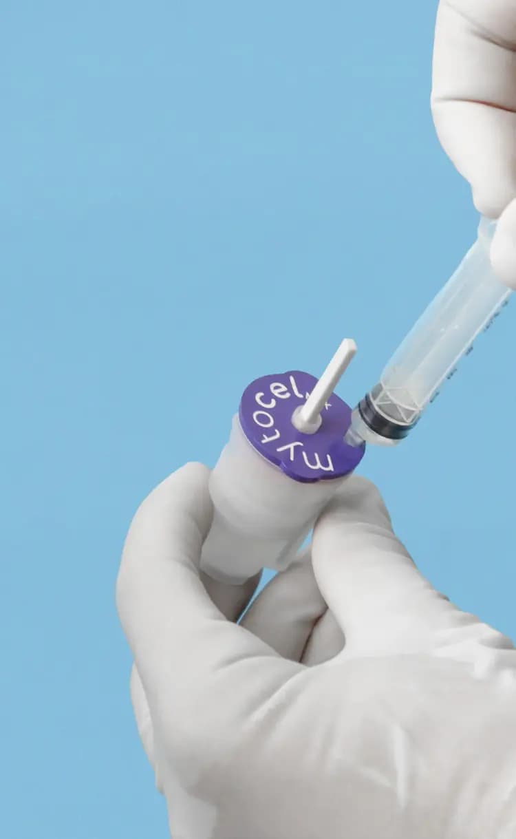
Mytocel MSK® is built on Rigenera® technology and the AMT® (Autologous Micrografting Technology) platform.
What is in the suspension
Cells, pericytes and matrix from the patient’s own cartilage.
Characterisation studies describe a heterogeneous suspension of cartilage-derived cells (chondrocytes and progenitor cells), pericytes and extracellular-matrix components from the donor tissue.1
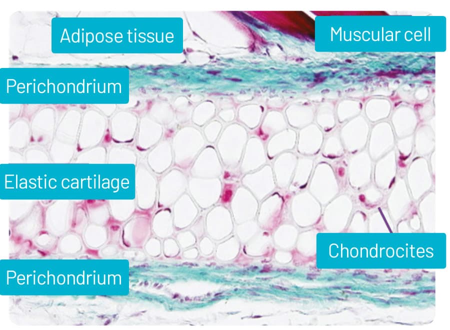
- Chondrocytes and progenitor cells
- Cartilage-derived cells from the donor tissue, as described in the characterisation studies.1
- Pericytes
- Carried from the donor tissue alongside the cartilage-derived cells, as described in the characterisation studies.1
- Extracellular-matrix components
- Matrix components from the donor tissue, collected in the same heterogeneous suspension as the cells.1
Processing
Mechanical, not biological.
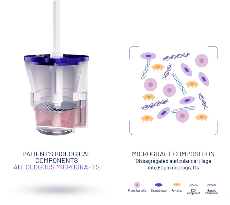
Mechanical disaggregation only: no laboratory expansion, no enzymatic digestion, no culture step.
Micrografts are collected through a calibrated 80 µm filter, the size range associated with viable progenitor-rich fractions in the characterisation work.1
Unlike cultured chondrocyte implantation, there is no laboratory expansion and no second procedure.
| Mytocel MSK | Cultured chondrocyte implantation | |
|---|---|---|
| Laboratory culture | None | Laboratory expansion |
| Number of procedures | One session | Two procedures |
| Tissue source | Patient’s own auricular cartilage | Patient’s own cartilage, expanded in a laboratory |
System components
Rigeneracons RS and the N4SA processing unit.
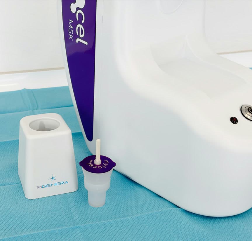
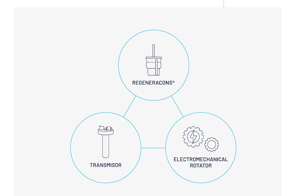
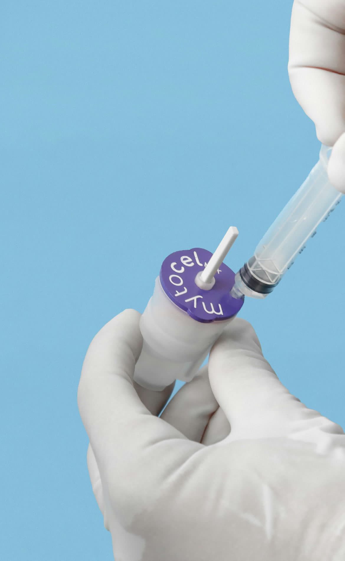
The single-use device carries the sample and its calibrated grid; the transmitter couples it to the processing unit; the unit’s electromechanical rotator drives the disaggregation cycle. No reagents and no culture step.
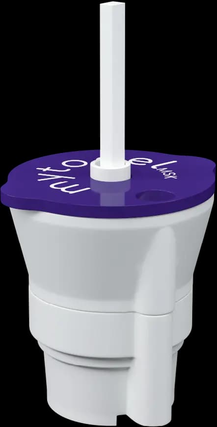
Rigeneracons RS
Disaggregation device
Rigeneracons® RS: sterile, single-use Class IIa medical device, designed to mechanically disaggregate the auricular cartilage sample and collect the micrograft suspension through the calibrated 80 µm filter.
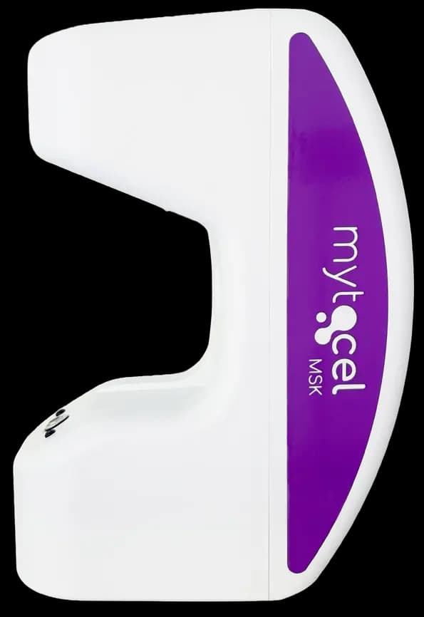
Rigenera N4SA
Processing unit
Rigenera® N4SA: Class I motor-driven processing unit. The reusable unit provides the calibrated rotational impulse for the documented processing cycle.
Storage. Store at room temperature in a dry, ventilated place.
Regulatory status
Where the evidence sits
Mechanistic rationale, pre-clinical models, small clinical series.
Nine publications and two preliminary reports: a three-year follow-up series (n = 8), two prospective ten-patient studies to 6 and 12 months, preliminary clinical reports, pre-clinical models and mechanistic characterisation. No randomised controlled trial has been published.234

References
- 1.Viganò M, Tessaro I, Trovato L, et al. Rationale and pre-clinical evidence for autologous cartilage micrografts in cartilage repair. 2018. View source
- 2.Marcarelli M, Zappia M, Rissolio L, et al. Cartilage micrografts as a novel non-invasive and non-arthroscopic autograft procedure for knee chondropathy: three-year follow-up. J Clin Med. 2021;10(2):322. View source
- 3.Tsoukas D, Muntean I, Simos C, Sabido-Vera R. Prospective observational study of a non-arthroscopic autologous cartilage micrografting technology for knee osteoarthritis. Bioengineering. 2023;10(11):1294. View source
- 4.Helito CP, Pessei V, Zaniboni C, Muntean I, et al. Efficacy of autologous micrografting technology in managing osteoarthritis pain: a pilot study. Bioengineering. 2024;11(11):1119. View source
Bring Mytocel MSK to your clinic.
UK stock, protocol training, first-case support and structured outcome collection, from the exclusive UK supplier.